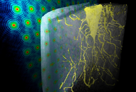
A new X-ray microscope technique gives a super-detailed look inside small samples, and does it in 3-D.
NEW VIEW A new X-ray microscopy technique reveals fine details inside a bone sample, such as holes housing bone cells. A detector measures changes in X-rays (depicted by colored dots, left) to create 2-D pictures that are combined into one 3-D image.
Log in
Subscribers, enter your e-mail address for full access to the Science News archives and digital editions.